

IMAGING RAMAN MICROSCOPE
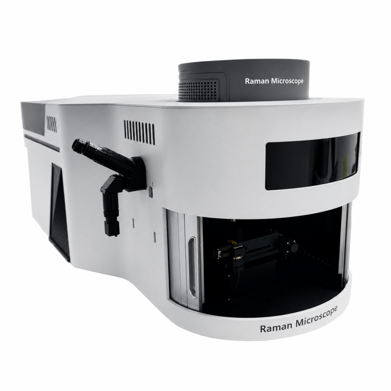
Raman Microscope
The Raman Microscope is a groundbreaking macro Raman spectral imaging instrument developed through more than 20 years of Raman spectrometer research and three years of dedicated product development. Its advanced optical design incorporates high-power lasers, linear laser shaping technology, high-spatial-resolution spectrometers, and an ultra-low-temperature cooled (-70°C) high-sensitivity CCD camera, enabling rapid scanning and high-resolution Raman imaging over large sample areas.
The system includes powerful multifunctional software that has been refined through extensive testing by hundreds of scientists worldwide. Continuous improvements based on user feedback have resulted in a highly stable, comprehensive, and user-friendly platform, making it an excellent solution for advanced Raman spectroscopy research and analysis.
Combining the capabilities of a high-performance optical microscope with a Raman spectrometer, the Raman Microscope provides a true “what you see is what you measure” experience. Its precision microscopic Raman detection platform enables accurate analysis of microscopic regions, significantly improving efficiency for micro-area investigations.
The advanced optical design minimizes optical path losses during camera imaging by separating the camera imaging path from the Raman signal collection path. This innovative approach maximizes Raman signal intensity while maintaining clear visual imaging for superior analytical performance.
Designed to operate reliably in demanding laboratory and industrial environments, the Raman Microscope is widely used in scientific research, food safety, pharmaceutical engineering, advanced materials research, nanotechnology, semiconductor analysis, forensic science, and new energy applications, providing fast, accurate, and non-destructive chemical characterization
specification
|
Confocal
Raman Microscope |
|
|
Excitation
Wavelength |
325、405、473、532、638、785、1064nm
selectable, up to 4 wavelengths can be integrated simultaneously |
|
Integration
Time |
8ms~1.3hours |
|
SNR |
>8000:1 |
|
Dynamic Range |
50000:1 |
|
Detector |
Ultra-low
temperature cooled, ultra-high sensitivity area CCD |
|
Spectral
Resolution |
2.0cm-1 |
|
Spot Diameter |
1mm |
|
Working
Humidity |
< 90% |
|
Working
Temperature |
10~35 ℃ |
|
Interface |
USB 3.0 |
|
Microscopic
Illumination |
High-brightness
long-life white LED |
|
Microscope
Camera |
5-megapixel
industrial camera |
|
Focus Method |
Conjugate
focusing |
|
Laser
Linewidth |
<0.2 nm |
|
Laser Power |
325nm 5 W
150~8000 2.6 405nm 5 W 150~8000 2.3 473nm 5 W 150~8000 2.1 532nm 30 W
150~8000 2.0 638nm 5 W 150~8000 2.0 785nm 30 W 150~8000 2.0 1064nm 30 W
150~8000 3.0 |
|
Voltage |
100~240 VAC |
|
Peak Power |
< 200 W |
|
Dimensions |
45×33×13.2
cm3 |
|
Weights |
14.5 Kg |
FEATURES
- 4
excitation wavelengths Optional
- Line-scan
Raman imaging
- Spatial
resolution up to 512 bands (single scan).
- Cooled
and high sensitivity detector.
- Cooled
to -70°C.
- Low
noise circuitry.
- Powerful
PC-based software.
- USB
3.0 interface.
- Automated
Raman imaging experiments with auto-focus and auto-scan.
- Long
focal length high-resolution design.
- Sealed
chamber design
- Large
imaging range (100x100mm) with automatic image stitching.
- Advanced
spectral processing algorithms and software.
- User-friendly
interface.