

BCF296 Laser Scanning Confocal Microscope
Confocal Microscope
The BCF296 is an all-new generation laser scanning confocal microscope with an industry-leading 25mm field of view and 8192*8192 pixel scanning, designed for high-throughput, high-quality imaging in modern biological research.
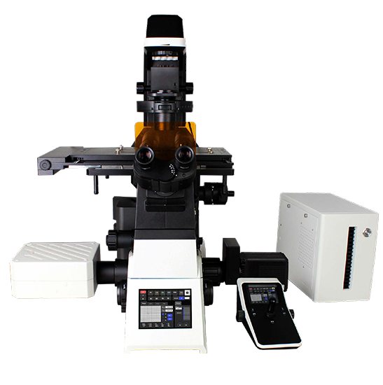
Description
As life science research demands deeper and broader analysis of tissues, organs and model organisms, efficient imaging solutions have become pivotal for scientific breakthroughs. Capturing images of such large samples requires expanding imaging ranges and reducing acquisition times to detect more accurate intracellular reactions. Following the legacy of its commercially successful predecessors, the BCF295 confocal microscopes, we proudly announce the triumphant upgrade completion of our all-new generation confocal microscope, the BCF296, specifically designed to meet the stringent requirements of modern biological research for high-throughput and high-quality imaging.
This upgrade not only marks significant advancements in optical systems, resolution, and acquisition speeds, but also introduces profound optimizations in intelligent operation and motorized design, transforming intricate experimental procedures into effortless tasks, thereby fulfilling the stringent demands of cutting-edge research across disciplines.
The BCF296 is equipped with an industry-leading 25mm field of view, effortlessly encompassing extensive sample areas for seamless large-scale imaging. With a scanning resolution of 8192*8192 pixels, it achieves vivid and precise imaging even at low magnifications. Its remarkable high-speed imaging capability of up to 60 fps (at 8*256 pixels) not only accommodates broad field observations but also delivers high-resolution imagery within a single frame, thereby gathering more data faster in each image.
Larger fields of view, enhanced resolution, and increased speed translate to superior image quality, clearer contrasts, and truer colors. This empowers you to delve deeper into the nanoscale universe within cells, interpreting cellular reaction processes more intuitively and accurately.
Model: BCF296
Laser: 405 nm, 488 nm, 561 nm, 640 nm
Detector: Standard detector, wavelength: 400-750nm, 4-channel PMT detector
Scanning Head: Maximum pixel size: 8192*8192
Eyepiece (Field of View): 10X, adjustable diopter -5 to +5
Objective: Apochromatic Objectives 10×, 20×, 40×, 60×
Condenser: 7-hole turret condenser with electric control, NA0.55, WD26. 7 positions for phase contrast, Hoffmann phase contrast, DIC, ND filter
Illumination System: Transmitted Kohler illumination, 3W LED
FEATURES
- To extract more data from the sample: industry-leading 25mm field of view captures large samples in a single scan with 1.5 times the data throughput of previous generations, at a scanning resolution up to 8192*8192 pixels.
- Fewer total images for large, high-resolution stitching: paired with the BS-2096 inverted microscope, it acquires high-quality 25 mm FOV confocal images, reducing the number of images and the acquisition time.
- High-performance objectives for confocal imaging: NIS series objectives with high numerical apertures, long working distances and exceptional chromatic aberration correction, including NIS Apochromat TIRF 100X Oil (NA up to 1.49), NIS Apochromat 20× C WI and the NIS Plan Apochromat series.
- High-efficiency scanning head and detectors: integrated high-precision scanning galvanometer system with a continuously variable electric pinhole for low noise, high contrast confocal imaging.
- Faster scanning speed: up to 60 frames per second at 8*256 pixels, reducing exposure time and phototoxicity and capturing dynamic events in real time.
- Real-time four-channel synchronized imaging: four-channel fluorescence merging with precise spectral unmixing for simultaneous detection of four fluorescent labels in the same field of view.
- Integrated software platform: high-speed hardware control, multi-dimensional imaging (X, Y, Z, λ and T scanning, multi-channel fluorescence, time-lapse, multi-position acquisition, Z-axis stacking and panoramic stitching) and 2D/3D deconvolution.
- High-performance BS-2096 platform: 25mm field of view with brightfield, fluorescence, differential interference contrast and phase contrast, plus single-layer or dual-layer optical path configurations.
- High-speed electric motor control: fast switching of objectives, filter blocks, XY stage and observation modules, with a joystick for intuitive stage control.
- Living cell culture system (optional): regulates temperature, humidity and CO₂ concentration for long-term live cell experiments.
- Adaptive Focus System (AFS): independent focusing design that eliminates focus drift for stable, sharp imaging, including super-resolution, confocal and TIRF applications.
- High specification LED fluorescent illumination: up to 4-channel LED illumination, instant-on, long service life and no bulb replacement, reducing photobleaching and phototoxicity.
- 5.6 inch touch screen with traditional knobs and buttons for intuitive control, even in dark laboratory environments.
- Large aperture observation optics and reflective fluorescence illuminator designed for the 25mm field of view, delivering high signal-to-noise ratio fluorescent images.
APPLICATION
- Life science research on tissues, organs and model organisms
- Large sample and high-throughput confocal imaging with large image stitching
- Live cell imaging and long-term observation of cellular dynamic processes
- Multi-channel fluorescence, Z-stack and time-lapse imaging of biological samples
Specifications
| OPTICAL SYSTEM | |
| Infinite optical system | ● |
| LASER | |
| Laser 405 nm, 488 nm, 561 nm, 640 nm | ● |
| DETECTOR | |
| Standard detector, wavelength: 400-750nm, 4-channel PMT detector | ● |
| Transmittance detector, single channel PMT detector | ○ |
| SCANNING HEAD | |
| Maximum pixel size: 8192*8192 | ● |
| SCAN MODE | |
| Supports combined use of X, Y, Z, λ and T scanning functions | ● |
| PINHOLE | |
| Electric stepless adjustment | ● |
| CONFOCAL FIELD OF VIEW | |
| Square inscribed in a Φ25mm diameter circle | ● |
| EYEPIECE | |
| 10X, adjustable diopter -5 to +5 | ● |
| VIEWING HEAD | |
| Siedentopf binocular viewing head, 10-40° inclined, Interpupillary 47-78mm, eyepiece interface Φ30mm. | ● |
| APOCHROMATIC PLAN OBJECTIVES | |
| 4X/ NA=0.16, WD=17.0mm, cover slip=0.17mm | ○ |
| 10X/ NA=0.45, WD=4.0mm, cover slip=0.17mm | ● |
| 20X/ NA=0.75, WD=1.1mm, cover slip=0.17mm | ● |
| (Wi) 20X/ NA=0.95, WD=1.1mm, cover slip=0.11-0.23mm | ○ |
| 40X/ NA=0.95, WD=0.21mm, cover slip=0.11-0.23mm | ● |
| (Oil) 60X/ NA=1.42, WD=0.25mm, cover slip=0.17mm | ● |
| (Oil) 100X/ NA=1.45, WD=0.13mm, cover slip=0.17 | ○ |
| (Oil) 100X/ NA=1.49, WD=0.09-0.16mm, cover slip=0.13-0.19mm (23°C), 0.14-0.20mm (37°C) | ○ |
| NOSEPIECE | |
| Motorized sextuple nosepiece with DIC expansion slot | ● |
| Motorized sextuple nosepiece with AFS module | ○ |
| STAGE | |
| Motorized control (grating type): Stage size: 445mm*300mm, travel range: 130mm*100mm, maximum speed: 25 mm/s. Positioning Accuracy: 0.1 μm. Repeatability: 0.5 μm. Equipped with a universal stage plate, compatible with 35mm-65mm culture dishes and slides, optional plate holder adapter available. | ● |
| CONDENSER | |
| 7-hole turret condenser with electric control, NA0.55, WD26. 7 positions for phase contrast, Hoffmann phase contrast, DIC, ND filter. | ● |
| FOCUSING SYSTEM | |
| Electric Z-axis, travel distance 10mm, precision 0.02μm, repeatability 0.1μm. Three-speed focusing knob: 2μm/revolution, 40μm/revolution, 200μm/revolution. | ● |
| ILLUMINATION SYSTEM | |
| Transmitted Kohler illumination, 3W LED | ● |
| Epi-Illumination: Widefield 4-band LED illumination. | ● |
| 6-Position electric fluorescence turntable (standard for B, G, U). | ● |
| Motorized Fluorescence Shutter. | ● |
| PORTS | |
| Beam splitter ratios: eyepiece 100% or left port 100% or right port 100% or eyepiece: right port=20:80. | ● |
| INTERMEDIATE MAGNIFICATION | |
| Manual 1X, 1.5X switching | ● |
| DIC PLATE | |
| 10X, 20X, 40X, 60X, 100X objective Inserts, compatible with the nosepiece turret | ○ |
| POWER CONTROL BOX | |
| X/Y/Z-axis electric control. Display objective magnification, fluorescence band and other states. Shortcut key settings. | ● |
| IMAGE BIT DEPTH | |
| 16 bits | ● |
| CAMERA | |
| Specialized camera | ● |
| SOFTWARE | |
| Featuring confocal scanning capabilities, multidimensional imaging, and pre-set channel functions, this software supports both wide-field and confocal imaging modes. It offers comprehensive hardware control modules, built-in image filtering tools, and supports export to various image formats, providing users with advanced imaging processing and management options. | ● |
| PC | |
| Computer workstation: a set of HP workstations or similar performance workstations. (1) HP Z840/CT Workstation/English OS Windows 7 64bit Professional Edition; (2) CPU: Intel Xeon E5-26434C 3.30 10MB 1600 x 1 or similar performance; (3) RAM: 32GB DDR -1600 ECC or similar performance; (4) HDD: 1TB 7200 RPM SATA 1ST HDD or similar performance; (5) 16X SuperMulti DVDRW SATA 1st ODD or similar performance; (6) Display: 2PCS ≥20 inch LED backlit widescreen IPS LCD displays. | ● |
| EXPANDABLE FUNCTIONS | |
| Live Cell Culturing System | ○ |
| Super-Resolution Structured Illumination Microscopy (SR-SIM) Module | ○ |
| Dual-Layer Fluorescence Heightened Accessor | ○ |
Note: ● Standard Outfit, ○ Optional